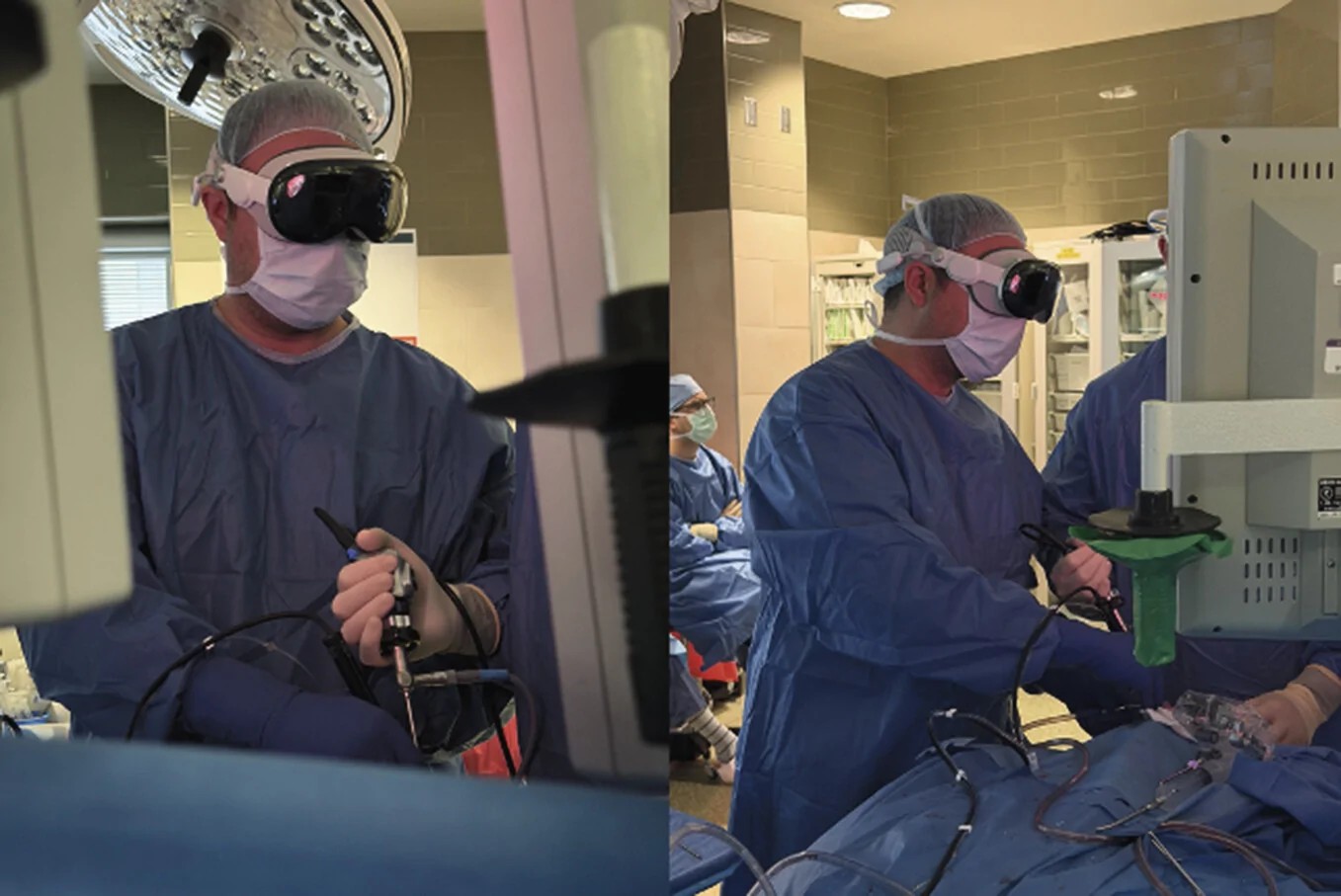
University of Pittsburgh surgeons have put a PEER-REVIEWED paper BEHIND a claim that circulated on social media this WEEK: the FIRST in-human skull base neurosurgery performed with an Apple Vision Pro on the surgeon’s head. The open-access case report in the Journal of Neurological Surgery Reports describes a PITUITARY tumor removed through the NOSE with NO complications. Read past the headline, though, and the paper is just as CANDID about what the headset did NOT do: it displayed ONE video FEED, ran on a BATTERY the team measured at roughly 90 MINUTES, and NEEDED more than an HOUR of SETUP.
What the paper reports
The report lists 13 AUTHORS from Pitt’s School of Medicine, UPMC neurosurgery and otolaryngology, Pitt’s School of Computing and Information, and CARNEGIE MELLON. It was received on June 16, accepted on August 25 and published online on October 6, according to the PubMed record and the article metadata. Neurosurgeon Georgios ZENONOS performed the neurosurgical portion; otolaryngologist Garret Choby’s team HARVESTED the nasoseptal flap and handled RECONSTRUCTION. The study ran under a university IRB protocol with WRITTEN patient consent.
The patient was a 38-year-old woman with Cushing disease and a 15-MILLIMETER hormone-secreting adenoma that, during surgery, was found to traverse the medial wall of the left cavernous sinus. The team drilled the sellar bone, EXPOSED both internal carotid arteries, and dissected the cavernous sinus wall to achieve COMPLETE resection. Postoperative MRI showed “no convincing residual pituitary mass.” She LEFT intensive care on day ONE, went home on day TWO, and showed NO cerebrospinal fluid leak at a day-five follow-up.
Pitt Neurosurgery flagged the publication on X on October 12:
Pitt surgeons have reported the first in-human skull base neurosurgery case performed with an Apple Vision Pro headset. @GAZenonosMD wore the headset while removing a pituitary tumor through the nose with otolaryngologist Garret Choby, MD. There were no complications.
— Pitt Neurosurgery (@PittNeurosurg) October 12, 2026
How the feed reached the headset
The ENGINEERING is CONVENTIONAL by DESIGN. An SDI cable carried the high-definition endoscope video from the camera control unit to “specialized capture hardware,” which COMPRESSED it for NETWORK transmission. The headset received it over a PRIVATE local network encrypted with WPA3, and the team MONITORED the router’s device list for unknown connections. The paper says the stream was EPHEMERAL: no patient images were retained on the capture box or on the Vision Pro. It does NOT name the capture hardware or the visionOS streaming app, which leaves OTHER hospitals UNABLE to REPRODUCE the stack from the text alone.
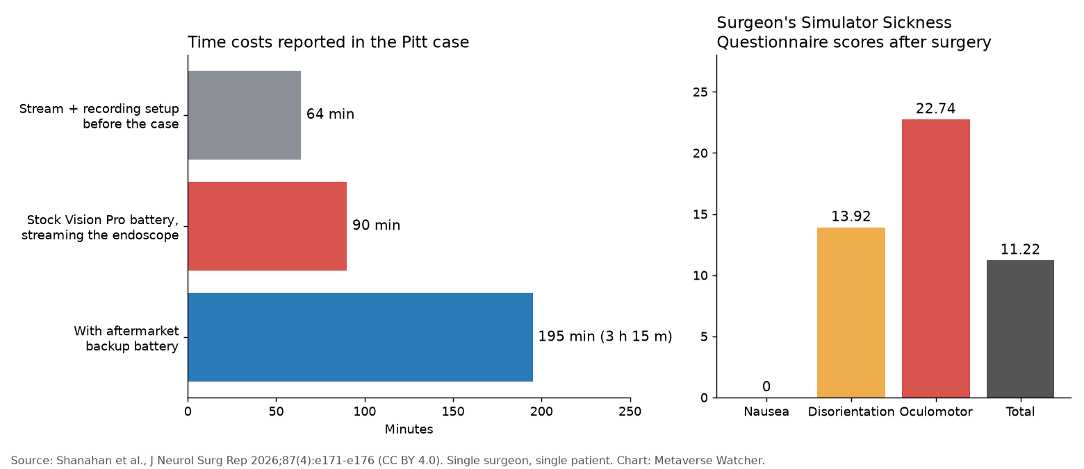
For the research record, a separate capture card wrote the endoscope output to a 256GB SD card, and a Blackmagic Studio Camera 4K Pro FILMED the room. Building the streaming and recording setup took 64 MINUTES, which the authors say happened ALONGSIDE routine preoperative preparation.
The surgeon put the headset on BEFORE scrubbing, with ZEISS prescription inserts fitted after an independent optometry check, then calibrated eye tracking and interpupillary distance using Apple’s standard flow. He ANCHORED the virtual screen where he wanted it and DECLINED a software option that lets the window follow head movement. Through passthrough he could STILL see the patient, cables, assistants and the room’s conventional monitors “without any clinically meaningful distortion,” the authors write.
The caveats sit in the paper itself
- ONE stream, by choice. A two-stream configuration adding Medtronic StealthStation navigation was AVAILABLE, and the paper’s Figure 2 shows it, but “for study purposes, only the endoscopic stream was displayed.” The otolaryngology team worked from the standard operating room monitor throughout.
- BATTERY is the binding limit. In pre-case tests streaming the endoscope, the STOCK Vision Pro battery lasted “roughly 90 min.” An AFTERMARKET backup stretched that to 3 HOURS 15 minutes, and the team designed a STERILE hot-swap procedure so the surgeon would not have to leave the field.
- NO latency number. The authors say video arrived “without perceptible delay, interruption, or system failure.” That is a SUBJECTIVE judgment; the paper publishes NO MEASURED latency.
- Some strain registered. The surgeon scored ZERO for nausea on the Simulator Sickness Questionnaire, but 13.92 for disorientation and 22.74 for oculomotor symptoms, for a total of 11.22. He completed ONLY the Likert portion of the SURG-TLX workload survey, scoring 26 of 120.
- N of ONE. The authors list the lack of a control group, subjective assessment, short follow-up and single-surgeon experience as LIMITATIONS, plus headset WEIGHT, NETWORK dependence and vergence-accommodation conflict. They declare no conflicts of interest and say a LARGER series is planned at Pitt.
The paper’s STRONGEST comparative evidence is NOT this case at all. It cites a 2026 Operative Neurosurgery study by Choi and colleagues in which skull base surgeons performed four CADAVERIC endonasal dissections and showed COMPARABLE error rates on FLAT monitors and mixed-reality streams.
An eight-month-old announcement, now with a headset name
Pitt first publicized MIXED-REALITY skull base surgery in a February 17, 2026 release describing a pituitary resection by Zenonos and Choby at UPMC Presbyterian. That release did NOT NAME a headset and spoke of bringing “key patient-specific information” into the surgeon’s working space. The new paper does NOT DATE its operation, so the two cannot be matched with CERTAINTY from the public record. What the paper DOES pin down is NARROWER: the information delivered in its case was the ENDOSCOPE feed alone.
The Metaverse Watcher take
This is a SAFETY and FEASIBILITY result, not a performance one, and the AUTHORS say so. Its VALUE for spatial computing is that it puts HARD OPERATING numbers on the RECORD: 90 MINUTES of stock battery, 64 MINUTES of setup, measurable OCULOMOTOR strain and an unnamed streaming stack. The authors call it a minimal “first feed.” The REAL test is the MULTI-STREAM dashboard they describe next, with NAVIGATION, VITALS and IMAGING in ONE view, and whether a larger series can show it HELPS surgeons rather than merely NOT HURTING patients.
Sources: Shanahan et al., Journal of Neurological Surgery Reports (PMC full text); publisher DOI; PubMed; Pitt Neurosurgery, February 17, 2026; @PittNeurosurg on X.